gALilEO CluB : eNJoy WitH Us


Thursday, August 19, 2010

:: AUTOPSY ::

What is an autopsy?
  • An autopsy (also known as a post-mortem examination or obduction) is the examination of the body of a dead person and is performed primarily to determine the cause of death, to identify or characterize the extent of disease states that the person may have had, or to determine whether a particular medical or surgical treatment has been effective. In academic institutions, autopsies sometimes are also requested for teaching and research purposes. Forensic autopsies are autopsies with legal implications and are performed to determine if death was an accident, homicide, suicide, or a natural event. The word autopsy is derived from the Greek word autopsia: "to see with one's own eyes."
  • Autopsies are performed by pathologists; medical doctors who have received specialty training in the diagnosis of diseases by the examination of body fluids and tissues.
Who determines whether an autopsy is performed?
  • A physician cannot order an autopsy on a patient without the consent of the next-of-kin. A medical examiner can order an autopsy without the consent of the next-of-kin. Deaths that are investigated by the medical examiner or coroner include all suspicious deaths, and, depending upon the jurisdiction, may include deaths of persons not being treated by a physician for a known medical condition, deaths of those who have been under medical care for less than 24 hours, or deaths that occurred during operations or other medical procedures.
  • In all other cases, consent must be obtained from the next-of-kin before an autopsy is performed, even at academic institutions or hospitals. The next-of-kin also has the right to limit the scope of the autopsy (for example, excluding the brain from evaluation or limiting the procedure to examination of the abdomen) if he/she wishes.

photo: Autopsy on Mussolini's corpse (16K)


View Image


Anatomical Male Autopsy Body with Complete Organs $2000

::SKIN CANCER::

Introduction of skin cancer

Skin cancer is the most common form of human cancer. It is estimated that over 1 million new cases occur annually. The annual rates of all forms of skin cancer are increasing each year, representing a growing public concern. It has also been estimated that nearly half of all Americans who live to age 65 will develop skin cancer at least once.
The most common warning sign of skin cancer is a change in the appearance of the skin, such as a new growth or a sore that will not heal.
The term "skin cancer" refers to three different conditions. From the least to the most dangerous, they are:
The two most common forms of skin cancer are basal cell carcinoma and squamous cell carcinoma. Together, these two are also referred to as nonmelanoma skin cancer. Melanoma is generally the most serious form of skin cancer because it tends to spread (metastasize) throughout the body quickly. Skin cancer is also known as skin neoplasia.
This article will discuss the two kinds of nonmelanoma skin cancer.

Basal cell carcinoma

What is basal cell carcinoma?
Basal cell carcinoma is the most common form of skin cancer and accounts for more than 90% of all skin cancer in the U.S. These cancers almost never spread (metastasize) to other parts of the body. They can, however, cause damage by growing and invading surrounding tissue.

Numerous Skin Cancer Pictures
Skin Cancer


What are risk factors for developing basal cell carcinoma?
Light-colored skin, sun exposure, and age are all important factors in the development of basal cell carcinomas. People who have fair skin and are older have higher rates of basal cell carcinoma. About 20% of these skin cancers, however, occur in areas that are not sun-exposed, such as the chest, back, arms, legs, and scalp. The face, however, remains the most common location for basal cell lesions. Weakening of the immune system, whether by disease or medication, can also promote the risk of developing basal cell carcinoma. Other risk factors include
  • exposure to sun. There is evidence that, in contrast to squamous cell carcinoma, basal cell carcinoma is promoted not by accumulated sun exposure but by intermittent sun exposure like that received during vacations, especially early in life. According to the U.S. National Institutes of Health, ultraviolet (UV) radiation from the sun is the main cause of skin cancer. The risk of developing skin cancer is also affected by where a person lives. People who live in areas that receive high levels of UV radiation from the sun are more likely to develop skin cancer. In the United States, for example, skin cancer is more common in Texas than it is in Minnesota, where the sun is not as strong. Worldwide, the highest rates of skin cancer are found in South Africa and Australia, which are areas that receive high amounts of UV radiation.
  • age. Most skin cancers appear after age 50, but the sun's damaging effects begin at an early age. Therefore, protection should start in childhood in order to prevent skin cancer later in life.
  • exposure to ultraviolet radiation in tanning booths. Tanning booths are very popular, especially among adolescents, and they even let people who live in cold climates radiate their skin year-round.
  • therapeutic radiation, such as that given for treating other forms of cancer. 
 What does basal cell carcinoma look like?

  • A basal cell carcinoma usually begins as a small, dome-shaped bump and is often covered by small, superficial blood vessels called telangiectases. The texture of such a spot is often shiny and translucent, sometimes referred to as "pearly." It is often hard to tell a basal cell carcinoma from a benign growth like a flesh-colored mole without performing a biopsy. Some basal cell carcinomas contain melanin pigment, making them look dark rather than shiny.
  • Superficial basal cell carcinomas often appear on the chest or back and look more like patches of raw, dry skin. They grow slowly over the course of months or years.
  • Basal cell carcinomas grow slowly, taking months or even years to become sizable. Although spread to other parts of the body (metastasis) is very rare, a basal cell carcinoma can damage and disfigure the eye, ear, or nose if it grows nearby.

How is basal cell carcinoma diagnosed?
  • To make a proper diagnosis, doctors usually remove all or part of the growth by performing a biopsy. This usually involves taking a sample by injecting a local anesthesia and scraping a small piece of skin. This method is referred to as a shave biopsy. The skin that is removed is then examined under a microscope to check for cancer cells.

How is basal cell carcinoma treated?
  • There are many ways to successfully treat a basal cell carcinoma with a good chance of success of 90% or more. The doctor's main goal is to remove or destroy the cancer completely with as small a scar as possible. To plan the best treatment for each patient, the doctor considers the location and size of the cancer, the risk of scarring, and the person's age, general health, and medical history.

Methods used to treat basal cell carcinomas include:
  • Curettage and desiccation: Dermatologists often prefer this method, which consists of scooping out the basal cell carcinoma by using a spoon like instrument called a curette. Desiccation is the additional application of an electric current to control bleeding and kill the remaining cancer cells. The skin heals without stitching. This technique is best suited for small cancers in non-crucial areas such as the trunk and extremities.
  • Surgical excision: The tumor is cut out and stitched up.
  • Radiation therapy: Doctors often use radiation treatments for skin cancer occurring in areas that are difficult to treat with surgery. Obtaining a good cosmetic result generally involves many treatment sessions, perhaps 25 to 30.
  • Cryosurgery: Some doctors trained in this technique achieve good results by freezing basal cell carcinomas. Typically, liquid nitrogen is applied to the growth to freeze and kill the abnormal cells.
  • Mohs micrographic surgery: Named for its pioneer, Dr. Frederic Mohs, this technique of removing skin cancer is better termed "microscopically controlled excision." The surgeon meticulously removes a small piece of the tumor and examines it under the microscope during surgery. This sequence of cutting and microscopic examination is repeated in a painstaking fashion so that the basal cell carcinoma can be mapped and taken out without having to estimate or guess the width and depth of the lesion. This method removes as little of the healthy normal tissue as possible. Cure rate is very high, exceeding 98%. Mohs micrographic surgery is preferred for large basal cell carcinomas, those that recur after previous treatment, or lesions affecting parts of the body where experience shows that recurrence is common after treatment by other methods. Such body parts include the scalp, forehead, ears, and the corners of the nose. In cases where large amounts of tissue need to be removed, the Mohs surgeon sometimes works with a plastic (reconstructive) surgeon to achieve the best possible postsurgical appearance.
  • Medical therapy using creams that attack cancer cells (5-Fluorouracil--5-FU, Efudex, Fluoroplex) or stimulate the immune system (imiquimod [Aldara]). These are applied several times a week for several weeks. They produce brisk inflammation and irritation. The advantages of this method is that it avoids surgery, lets the patient perform treatment at home, and may give a better cosmetic result. Disadvantages include discomfort, which may be severe, and a lower cure rate, which makes medical treatment unsuitable for treating most skin cancers on the face.
How is basal cell carcinoma prevented?
Avoiding sun exposure in susceptible individuals is the best way to lower the risk for all types of skin cancer. Regular surveillance of susceptible individuals, both by self-examination and regular physical examination, is also a good idea for people at higher risk. People who have already had any form of skin cancer should have regular medical checkups.
Common sense preventive techniques include
  • limiting recreational sun exposure;
  • avoiding unprotected exposure to the sun during peak radiation times (the hours surrounding noon);
  • wearing broad-brimmed hats and tightly-woven protective clothing while outdoors in the sun;
  • regularly using a waterproof or water resistant sunscreen with UVA protection and SPF 30 or higher;
  • undergoing regular checkups and bringing any suspicious-looking or changing lesions to the attention of the doctor; and
  • avoiding the use of tanning beds and using a sunscreen with an SPF of 30 and protection against UVA (long waves of ultraviolet light.). Many people go out of their way to get an artificial tan before they leave for a sunny vacation, because they want to get a "base coat" to prevent sun damage. Even those who are capable of getting a tan, however, only get protection to the level of SPF 6, whereas the desired level is an SPF of 30. Those who only freckle get little or no protection at all from attempting to tan; they just increase sun damage. Sunscreen must be applied liberally and reapplied every two to three hours, especially after swimming or physical activity that promotes perspiration, which can weaken even sunscreens labeled as "waterproof."

 Gallery of Skin Cancer Pictures

  • Excessive exposure to sunlight is the main cause of skin cancer. Sunlight contains ultraviolet (UV) rays that can alter the genetic material in skin cells, causing mutations. Sunlamps, tanning booths, and X-rays also generate UV rays that can damage skin and cause malignant cell mutations.
  • From the least to most dangerous, skin cancer refers to three different conditions: basal cell carcinoma, squamous cell carcinoma (the first stage of which is called actinic keratosis) and melanoma. The two most common forms of skin cancer are basal cell carcinoma and squamous cell carcinoma. Together, these two are also referred to as nonmelanoma skin cancer. Melanoma is generally the most serious form of skin cancer because it tends to spread (metastasize) throughout the body quickly.


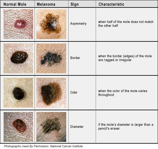
 cancer-moles.gif (108754 bytes)


:: ectopic pregnancy::

DEFINITION
  • Ectopic means "out of place." In an ectopic pregnancy, a fertilized egg has implanted outside the uterus. The egg settles in the fallopian tubes in more than 95% of ectopic pregnancies. This is why ectopic pregnancies are commonly called "tubal pregnancies." The egg can also implant in the ovary, abdomen, or the cervix, so you may see these referred to as cervical or abdominal pregnancies.
  • None of these areas has as much space or nurturing tissue as a uterus for a pregnancy to develop. As the fetus grows, it will eventually burst the organ that contains it. This can cause severe bleeding and endanger the mother's life. A classical ectopic pregnancy does not develop into a live birth.

Signs and Symptoms
  • Ectopic pregnancy can be difficult to diagnose because symptoms often mirror those of a normal early pregnancy. These can include missed periods, breast tenderness, nausea, vomiting, or frequent urination.
  • The first warning signs of an ectopic pregnancy are often pain or vaginal bleeding. You might feel pain in your pelvis, abdomen, or, in extreme cases, even your shoulder or neck (if blood from a ruptured ectopic pregnancy builds up and irritates certain nerves). Most women describe the pain as sharp and stabbing. It may concentrate on one side of the pelvis and come and go or vary in intensity.
  • Any of the following additional symptoms can also suggest an ectopic pregnancy:
*  vaginal spotting
*  dizziness or fainting (caused by blood loss)
*  low blood pressure (also caused by blood loss)
*  lower back pain
 

 ectopic pregnancy pathophysiology
the picture shows about the ectopic pregnancy. the fetus at the ovary not at the uterus,

::shyphilis in BOYS::

What are some signs of shyphilis in boys?

  • There are different manifestation of syphilis, depending on the the stage of the disease - primary, secondary tertiary, neurosyphilis and resting one. Primary syphilis is typically acquired via direct sexual contact near the infectious lesions of a character with syphilis. 
  • Contrary to popular belief, syphilis can not be contracted through toilet chairs, daily endeavours, hot tubs, or sharing eating utensils or clothing.
  • Approximately 10-90 days after the initial exposure (average 21 days), a skin lesion may be see on the genitalia. This lesion, called a chancre, is a firm, painless skin ulceration localized at the point of initial exposure to the spirochete, recurrently on the penis, vagina or rectum. Rarely, there may be multiple lesion present although typically only one lesion is see. The lesion may persist for 4 to 6 weeks and usually heal spontaneously. Local lymph node swelling can occur. During the initial incubation extent, individuals are otherwise asymptomatic..
  • .but read here for more:
  • http://en.wikipedia.org/wiki/syphilis...


SYMPTOMS
  • Main symtom is a generalized rash on the trunk of the body and clear leisions on the shaft of the penis!


Many types of syphilis
(1)Primary syphilis is typically acquired via direct sexual contact beside the infectious lesions of a individual with syphilis. Approximately 10-90 days after the initial exposure (average 21 days), a skin lesion may be see on the genitalia. This lesion, called a chancre, is a firm, painless skin ulceration localized at the point of initial exposure to the spirochete, commonly on the penis, vagina or rectum. Rarely, there may be multiple lesion present although typically only one lesion is see. The lesion may persist for 4 to 6 weeks and usually heal spontaneously. Local lymph node swelling can occur. During the initial incubation term, individuals are otherwise asymptomatic. As a result, many patients do not want medical care straight.

(2)Secondary syphilis occurs approximately 1-6 months (commonly 6 to 8 weeks) after the primary infection. There are tons different manifestations of lower disease. There may be a symmetrical reddish-pink non-itchy rash on the trunk and extremities.The reckless can involve the palms of the hands and the soles of the foot. In moist areas of the body, the rash become flat broad whitish lesions prearranged as condylomata lata. Mucous patches may also appear on the genitals or contained by the mouth. All of these lesions are infectious and harbor live treponeme organisms. A patient beside syphilis is most contagious when he or she has lesser syphilis. Other symptoms common at this stage include hallucination, sore throat, malaise, weight loss, headache, meningismus, and enlarged lymph nodes. Rare manifestation include an acute meningitis that occurs within about 2% of patients, hepatitis, renal disease, hypertrophic gastritis, patchy proctitis, ulcerative colitis, rectosigmoid mass, arthritis, periostitis, optic neuritis, intersitial keratitis, iritis, and uveitis

(3)Latent syphilis is defined as have serologic proof of infection without signs or symptoms of disease. Latent syphilis is further described as any early or postponed. Early latent syphilis is defined as have syphilis for two years or less from the time of initial infection lacking signs or symptoms of disease. Late latent syphilis is infection for greater than two years but in need clinical evidence of disease. The distinction is important for both analysis and risk for transmission. In the real-world, the timing of infection is normally not known and should be presumed to be behind for the purpose of therapy. Early resting syphilis may be treated with a single intramuscular injection of a long-acting penicillin. Late sleeping syphilis, however, requires three weekly injections. For infectiousness, however, late resting syphilis is not considered as contagious as early inactive syphilis.


(4)Tertiary syphilis usually occurs 1-10 years after the initial infection, though in some cases it can pinch up to 50 years. This stage is characterized by the formation of gummas which are soft, tumor-like balls of inflammation agreed as granulomas. The granulomas are chronic and represent an inability of the immune system to completely clear the organism. Gummas were once readily see in the skin and mucous membranes although they tend to transpire internally in recent history. They may appear almost anywhere in the body including in the skeleton. The gummas produce a chronic inflammatory state in the body with mass-effects upon the local anatomy. Other characteristics of untreated tertiary syphilis include neuropathic mutual disease, which are a degeneration of joint surfaces resulting from loss of sensation and fine position sense (proprioception).


 Go to fullsize image


 Go to fullsize image

::glaucoma::

Glaucoma and Your Eyes
  • Glaucoma is an eye condition that develops when too much fluid pressure builds up inside of the eye. It tends to be inherited and may not show up until later in life.
  • The increased pressure, called intraocular pressure, can damage the optic nerve, which transmits images to the brain. If damage to the optic nerve from high eye pressure continues, glaucoma will cause loss of vision. Without treatment, glaucoma can cause total permanent blindness within a few years.
  • Because most people with glaucoma have no early symptoms or pain from this increased pressure, it is important to see your ophthalmologist regularly so that glaucoma can be diagnosed and treated before long-term visual loss occurs.
  • If you are over the age of 40 and if you have a family history of glaucoma, you should have a complete eye exam with an ophthalmologist every one to two years. If you have health problems such as diabetes or a family history of glaucoma or are at risk for other eye diseases, you may need to visit your eye doctor more frequently.


Why Does Pressure Rise in the Eye to Cause 

Glaucoma?

  • Glaucoma usually occurs when intraocular pressure increases. This happens when the fluid pressure in the eye's anterior chamber, the area between the cornea and the iris, rises.
  • Normally, this fluid, called aqueous humor, flows out of the eye through a mesh-like channel. If this channel becomes blocked, fluid builds up, causing glaucoma. The direct cause of this blockage is unknown, but doctors do know that it is most often inherited, meaning it is passed from parents to children.
  • Less common causes of glaucoma include a blunt or chemical injury to the eye, severe eye infection, blockage of blood vessels in the eye, inflammatory conditions of the eye, and occasionally eye surgery to correct another condition. Glaucoma usually occurs in both eyes, but it may involve each eye to a different extent.

What Are the Types of Glaucoma?

  • There are two main types of glaucoma:

1.     Open-angle glaucoma.
Also called wide-angle glaucoma, this is the most common type of glaucoma. The structures of the eye appear normal, but fluid in the eye does not flow properly through the drain of the eye, called the trabecular meshwork.


2.     Angle-closure glaucoma. 
Also called acute or chronic angle-closure or narrow-angle glaucoma, this type of glaucoma is less common, but can cause a sudden buildup of pressure in the eye. Drainage may be poor because the angle between the iris and the cornea (where a drainage channel for the eye is located) is too narrow. Or, the pupil opens too wide, narrowing the angle and blocking the flow of the fluid through that channel.


Who Gets Glaucoma?

  • Glaucoma most often occurs in adults over age 40, but it can also occur in young adults, children, and even infants. In African-Americans, glaucoma occurs more frequently and at an earlier age and with greater loss of vision.
  • You are at an increased risk of glaucoma if you:

  1. Are of African-American, Irish, Russian, Japanese, Hispanic, Inuit, oScandinavian descent.
  2. Are over age 40.
  3. Have a family history of glaucoma.
  4. Have poor vision.
  5. Have diabetes.
  6. Take systemic corticosteroid medications, such as prednisone. 


Common Eye Problems

:: vaginal prolapsed ::

VAGINAL PROLAPSE
  •  A vaginal prolapse is a condition in which structures such as the uterus, rectum, bladder, urethra, small bowel, or the vagina itself may begin to prolapse, or fall, out of their normal positions. Without medical treatment or surgery, these structures may eventually prolapse farther and farther into the vagina or even through the vaginal opening if their supports weaken enough. 
  • The symptoms that result from vaginal prolapse commonly affect sexual functions and bodily functions such as urination and defecation. Pelvic pressure and discomfort are also common symptoms. 
  • The following are types of vaginal prolapse:
  1. *       Rectocele (prolapse of the rectum) - This type of vaginal prolapse involves a prolapse of the back wall of the vagina (rectovaginal fascia). When this wall weakens, the rectal wall pushes against the vaginal wall, creating a bulge. This bulge may become especially noticeable during bowel movements.
  2. *       Cystocele (prolapse of the bladder, bladder drop) - This can occur when the front wall of the vagina (pubocervical fascia) prolapses. As a result, the bladder may prolapse into the vagina. When this condition occurs, the urethra usually prolapses as well. A urethral prolapse is also called a urethrocele. When both the bladder and urethra prolapse, this condition is known as a cystourethrocele. Urinary stress incontinence (urine leakage during coughing, sneezing, exercise, etc) is a common symptom of this condition.
  3. *       Enterocele (herniated small bowel) - The weakening of the upper vaginal supports can cause this type of vaginal prolapse. This condition primarily occurs following a hysterectomy. An enterocele results when the front and back walls of the vagina separate, allowing the intestines to push against the vaginal skin.
  4. *       Prolapsed uterus (womb) - This involves a weakening of a group of ligaments called the uterosacral ligaments at the top of the vagina. This causes the uterus to fall, which commonly causes both the front and back walls of the vagina to weaken as well. The following are stages of uterine prolapse:


    o    First-degree prolapse: The uterus droops into the upper portion of the      vagina.


    o    Second-degree prolapse: The uterus falls into the lower part of the vagina.


    o    Third-degree prolapse: The cervix, which is located at the bottom of the uterus, sags to the vaginal opening and may protrude outside the body. This condition is also called procidentia, or complete prolapse.


    o    Fourth-degree prolapse: The entire uterus protrudes entirely outside the vagina. This condition is also called procidentia, or complete prolapse.

    • *       Vaginal vault prolapse - This type of prolapse may occur following a hysterectomy, which involves the removal of the uterus. Because the uterus provides support for the top of the vagina, this condition is common after a hysterectomy, with upwards of 10% of women developing a vaginal vault prolapse after undergoing a hysterectomy. In vaginal vault prolapse, the top of the vagina gradually falls toward the vaginal opening. This may cause the walls of the vagina to weaken as well. Eventually, the top of the vagina may protrude out of the body through the vaginal opening, effectively turning the vagina inside out. A vaginal vault prolapse often accompanies an enterocele.
    • Approximately 30-40% of women develop some presentation of vaginal prolapse in their lifetime, usually following menopause, childbirth, or a hysterectomy. Most women who develop this condition are older than 40 years. Many women who develop symptoms of a vaginal prolapse do not seek medical help because of embarrassment or other reasons. Some women who develop a vaginal prolapse do not experience symptoms.

    View Image

    View Image

    ::RENAL OSTEODYSTROPHY::

    What is renal osteodystrophy?

    • The medical term "renal" describes things related to the kidneys. Renal osteodystrophy is a bone disease that occurs when your kidneys fail to maintain the proper levels of calcium and phosphorus in your blood. It's a common problem in people with kidney disease and affects 90 percent of dialysis patients.
    • Renal osteodystrophy is most serious in children because their bones are still growing. The condition slows bone growth and causes deformities. One such deformity occurs when the legs bend inward toward each other or outward away from each other; this deformity is referred to as "renal rickets." Another important consequence is short stature. Symptoms can be seen in growing children with renal disease even before they start dialysis.
    • The bone changes from renal osteodystrophy can begin many years before symptoms appear in adults with kidney disease. For this reason, it's called the "silent crippler." The symptoms of renal osteodystrophy aren't usually seen in adults until they have been on dialysis for several years. Older patients and women who have gone through menopause are at greater risk for this disease because they're already vulnerable to osteoporosis, another bone disease, even without kidney disease. If left untreated, the bones gradually become thin and weak, and a person with renal osteodystrophy may begin to feel bone and joint pain. There's also an increased risk of bone fractures.
    Hormones and Minerals
    • In healthy adults, bone tissue is continually being remodeled and rebuilt. The kidneys play an important role in maintaining healthy bone mass and structure because one of their jobs is to balance calcium and phosphorus levels in the blood.
    • Calcium is a mineral that builds and strengthens bones. It's found in many foods, particularly milk and other dairy products. If calcium levels in the blood become too low, four small glands in the neck called the parathyroid glands release a hormone called parathyroid hormone (PTH). This hormone draws calcium from the bones to raise blood calcium levels. Too much PTH in the blood will remove too much calcium from the bones; over time, the constant removal of calcium weakens the bones.
    • Phosphorus, which is found in most foods, also helps regulate calcium levels in the bones. Healthy kidneys remove excess phosphorus from the blood. When the kidneys stop working normally, phosphorus levels in the blood can become too high, leading to lower levels of calcium in the blood and resulting in the loss of calcium from the bones.
    • Healthy kidneys produce calcitriol, a form of vitamin D, to help the body absorb dietary calcium into the blood and the bones. If calcitriol levels drop too low, PTH levels increase, and calcium is removed from the bones. Calcitriol and PTH work together to keep calcium balance normal and bones healthy. In a patient with kidney failure, the kidneys stop making calcitriol. The body then can't absorb calcium from food and starts removing it from the bones.
    How is renal osteodystrophy diagnosed?
    • To diagnose renal osteodystrophy, your doctor may take a sample of your blood to measure levels of calcium, phosphorus, PTH, and calcitriol. The doctor may perform a bone biopsy to see how dense your bones are. A bone biopsy is done under local anesthesia and involves removing a small sample of bone from the hip and analyzing it under a microscope. Determining the cause of renal osteodystrophy helps the doctor decide on a course of treatment.
    How is osteodystrophy treated?
    • Controlling PTH levels prevents calcium from being withdrawn from the bones. Usually, overactive parathyroid glands are controllable with a change in diet, dialysis treatment, or medication. The drug cinacalcet hydrochloride (Sensipar), approved by the Food and Drug Administration in 2004, lowers PTH levels by imitating calcium. If PTH levels can't be controlled, the parathyroid glands may need to be removed surgically.
    • If your kidneys aren't making adequate amounts of calcitriol, you can take synthetic calcitriol as a pill or in an injectable form. Your doctor may prescribe a calcium supplement in addition to calcitriol.
    • Renal osteodystrophy can also be treated with changes in diet. Reducing dietary intake of phosphorus is one of the most important steps in preventing bone disease. Almost all foods contain phosphorus, but it's especially high in milk, cheese, dried beans, peas, nuts, and peanut butter. Limit drinks such as cocoa, dark sodas, and beer. Often, medications such as calcium carbonate (Tums), calcium acetate (PhosLo), sevelamer hydrochloride (Renagel), or lanthanum carbonate (Fosrenol) are prescribed with meals and snacks to bind phosphorus in the bowel. These decrease the absorption of phosphorus into the blood. Be sure your phosphate binder is aluminum-free because aluminum can be toxic and cause anemia. A renal dietitian can help develop a dietary plan to control phosphorus levels in the blood.
    • Exercise has been found to increase bone strength in some patients. It's important, however, to consult a doctor or health care professional before beginning any exercise program.
    • A good treatment program, including proper attention to dialysis, diet, and medications, can improve your body's ability to repair bones damaged by renal osteodystrophy